Neuroendocrine Integration

Part Of: [Neuroanatomy] sequence

Table Of Contents

  • Circulatory System and BBB
  • Ventricular System and BCSFB
  • Endocrine System and CVOs
  • Visceral vs Cognitive Processing
  • The Folly Of Spock

Circulatory System and BBB

Cells need blood to survive. Neurons are cells like any other. Thus, we expect the circulatory system to extend into the brain.

Blood flow in the brain is organized around a structure poetically named the Circle Of Willis.

Neuroendocrine- Circle Of Willis

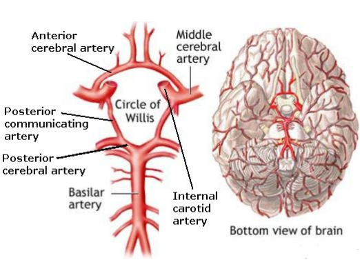
Unfortunately, capillaries sometimes leak, releasing noxious bacteria. Such infections are usually addressed by your immune system. However, certain areas of the body cannot tolerate such risks. If you had to choose, which areas of the body which you want special protection from infection?

Natural selection has produced specialized protection for three areas of the body: the brain, the eyes, the gonads.  Damage to these structures are particularly destructive to an organism’s fitness. Capillaries in these “protected zones” are given essentially several extra layers of “armor”, which prevent bacteria from escaping.

Neuroendocrine- BBB

This protective mechanism is known as the blood-brain barrier (BBB). This armor is metabolically expensive, which explains the absence of “blood-foot barriers”.

Ventricular System and BCSFB

Your brain does not rest against the base of your skull. That would destroy brain tissue. Instead, it is immersed in a fluid bath, which protects and supports the brain.  Formally, the fluid is known as cerebrospinal fluid (CSF), and it is found as a support for the brain (ventricular system) and surrounding/cushioning the brain (in the subarachnoid space).

Neuroendocrine- Ventricles and CSF

Cerebrospinal fluid (CSF) takes no direct connection to the circulatory system. In fact, since the CSF directly contacts nervous tissue, it also requires extra protection from infection. The blood-cerebrospinal fluid barrier (BCSFB) exists for precisely this reason.

Taken together, the BBB and the BCSFB insulate the brain from the circulatory system, while still allowing nutrients to reach neurons:

Neuroendocrine- BBB and BCSFB

Endocrine System and CVOs

In Towards Body Architecture we explored how the nervous system interacts with other bodily systems (respiratory, digestive, etc).  In fact, the nervous system acts as a control system, influencing how other systems perform. No other anatomical system can claim that…

…except one. Your endocrine system also acts as a control system! Hormones play a role in digestion, cell growth, reproduction, etc – a whole host of bodily processes. Speaking generally, nervous signals act quickly (nerve signals do their work within seconds), whereas endocrine control (hormone signals enact change more slowly, usually in minutes or hours).

Two independent control systems is a poor way to design a body. We have every reason to benefit from them synchronizing their efforts, exchanging information. Let’s examine stress as an example, which requires the involvement of both systems.

  • The nervous system is responsible for detecting stressful stimuli. It then transmits a signal, via nerves of the sympathetic nervous system to the adrenal gland, which immediately begins manufacturing cortisol (a hormone which mediates stress). This is fight-or-flight, a fast response.
  • The endocrine system concurrently releases adrenocorticotropic hormone (ACTH) which also prompts the adrenal gland to manufacture cortisol. However, ACTH takes longer to take effect, and stays in your system longer. Thus, the endocrine system supports non-transient forms of stress.

These two phenomena are obviously correlated. But how? Your endocrine system is confined to your circulatory system – hormones travel via the bloodstream. But we just learned that the brain is insulated from the bloodstream…

The nervous system and the endocrine system coordinate with one another via circumventricular organs (CVOs). CVOs puncture the blood-brain barrier in a controlled way. They come in two flavors:

  • Sensory CVOs translate hormone messengers into neural signals.
  • Secretory CVOs translate neural signals into hormone messengers.

As you might expect by now, these two categories of CVOs provide neuroendocrine integration.

Neuroendocrine- Visceral Loop

Visceral vs Cognitive Processing

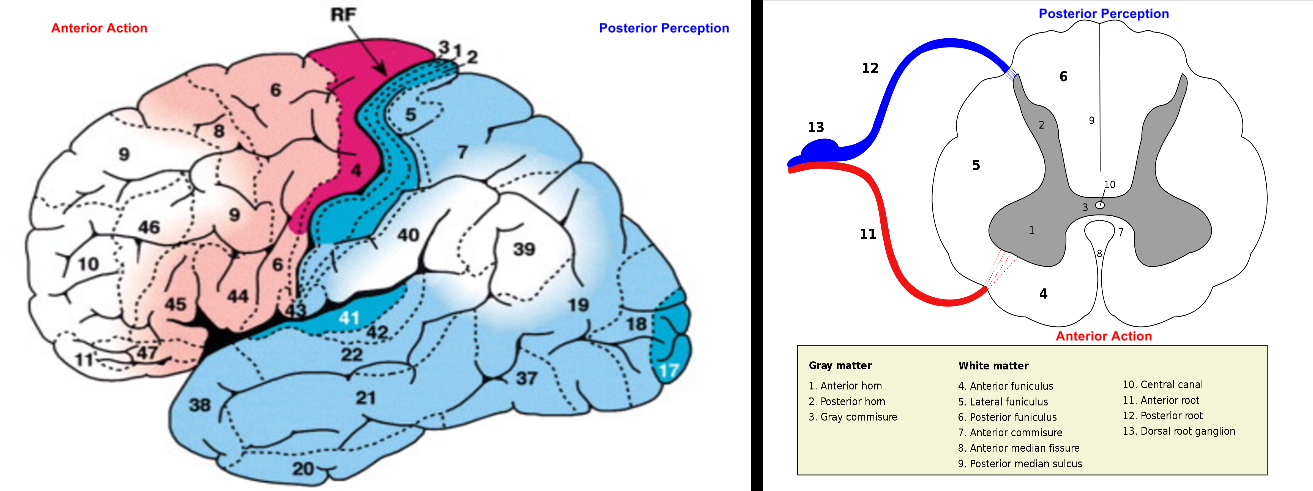
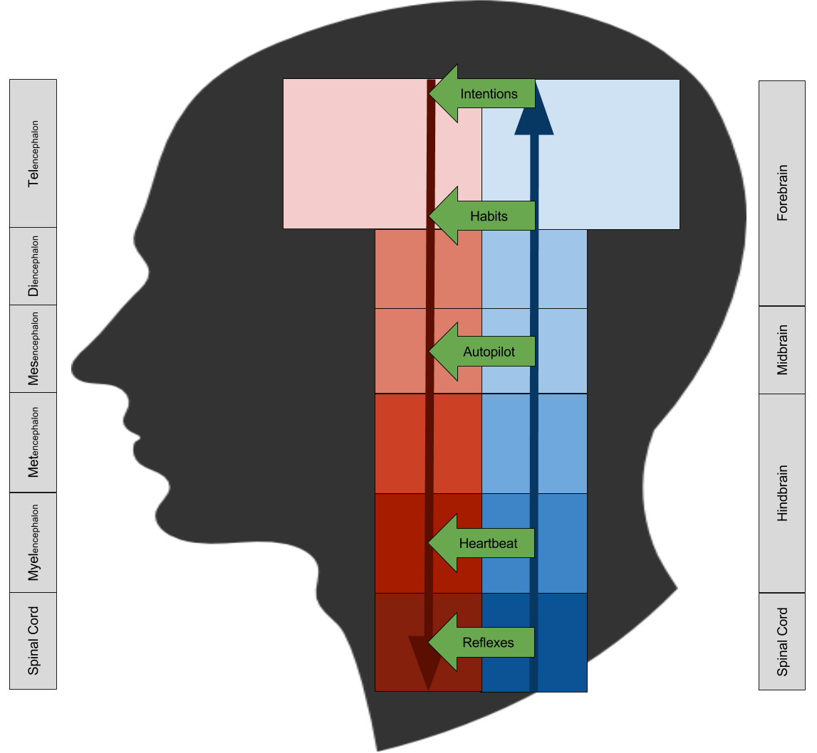
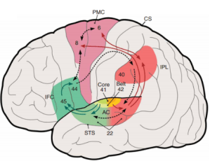
In my last post, I presented the cognitive perception-action cycle. Let us consider these cybernetic systems side by side.

Neuroendocrine- Abstraction Hierarchy

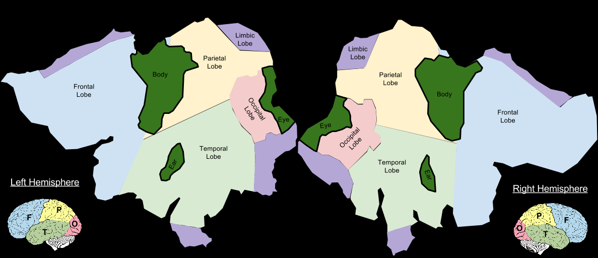
This image situates visceral and cognitive processes together. This reflects the medial viscera principle:

Visceral processes tend to reside in the center of the brain (medial regions).

The neuroendocrine system does not operate alone. Rather, it works alongside the autonomic nervous system, which regulates the body via two complementary systems:

  1. Sympathetic Nervous System, which promotes “fight for flight” readiness.
  2. Parasympathetic Nervous System, with a restorative “rest or digest” function.

In this way, the visceral perception-action cycle has two arms:

  1. The autonomic nervous system, which quickly perceives and regulates the body.
  2. The neuroendocrine system, which operates at a slower, more deliberate pace.

We can call the visceral perception-action cycle the “hot loop”, in contrast with the cognitive cycle, or “cold loop”. These loops represent a central organizing principle of the nervous system.

neuroendocrine-information-channels-2


The spinal cord bears nerve fibers in service of both loops, both autonomic nerves bearing sympathetic & parasympathetic signals, and somatic nerves which regulate the skin and musculature. The cranial nerves include exteroceptive nerves as well (vision, hearing, smell, etc) which together encode information about the external world. 

The Folly Of Spock

As we will see later, the visceral perception-action cycle participates in the neural basis of emotion.

Next time you watch Star Trek, you may safely infer the Spock’s species has not achieved neuroendocrine integration.

Emotional processes reside at the center of the brain. Emotion is thus ideally situated to modify, modulate, and alter your decision-making capabilities.

This tension between visceral and cognitive, between hot and cold, is one of the hallmarks of being human.

Neuroendocrine- Song Of Ice and Fire

Until next time.


Abstraction Hierarchy: Perception-Action Cycles

Part Of: [Neuroanatomy] sequence
Next Up: The Interlocking Loop Hypothesis

Posterior Perception, Anterior Action

Consider again the neural tube. In Brain Ontogeny, we learned how brains emerge from five little bumps in the neural tube.

Brain Ontogeny- Vesicle Differentiation Chronology

The neural tube develops under several organizing principles. One of the most important is the Bell-Magendie law: in the nervous system, motor nerves & computations happen in the front of an animal (anterior action), perceptual nerves & computations are at the back (posterior perception). This principle applies to the entire nervous system, from the spinal cord to the neocortex.

Cybernetics- Posterior Perception, Anterior Action

From an evolutionary vantage point, this principle is fairly obvious. Because gravity is a thing, the vertebrate body plan features anterior limbs (movement requires limbs between body & ground) and posterior vision (because of the sun’s position relative to the Earth). Notice how bipedalism and its deformation of the human neuraxis obscures this principle. Our quadruped ancestry thereby illuminates the functional organization of the human nervous system.

The Abstraction Hierarchy

The nervous system is a complex organ. Sometimes, choices do not require much analysis. Often, however, a decision becomes clear only after contemplation.

As information travels through the brain, it becomes more abstract, and resultant behavior more flexible. This is the brain’s abstraction hierarchy.

Cybernetics- Abstraction Hierarchy

Darker colors denote concrete representations, lighter colors denote abstraction.

In cognitive science, the following synonyms hold:

  • Information is a synonym for memory.
  • Sites of information processing is a synonym for modules.

The abstraction hierarchy can inform both levels of analyses. Let’s see an example.

Cognitive scientists like to distinguish between information encoding events (episodic memory) and information encoding concepts (semantic memory). The abstraction hierarchy explains the relation between the two: the concept of apple generalizes any number of events that contain apples. That is, semantic memory is situated at a higher level than episodic memory.

How do we know the brain is organized hierarchically? Brain damage provides one line of evidence.

  • Severe damage to the brain stem is fatal.
  • However, if a human loses everything but the brain stem, they cling to life (persistent vegetative state)
  • If the midbrain is spared, such that a person loses only their cerebrum, they can spontaneously move, but their behavior is disorganized.

Renowned neuroscientist Charles Sherrington once described the brain as an enchanted loom:

The brain is waking and with it the mind is returning. It is as if the Milky Way entered upon some cosmic dance. Swiftly the head mass becomes an enchanted loom where millions of flashing shuttles weave a dissolving pattern, always a meaningful pattern though never an abiding one; a shifting harmony of subpatterns.

The abstraction hierarchy teaches us: the higher you climb the enchanted loom, the more flexible your dance.

Biological Decision Making

We are now in a position to understand decision making.

Decisions are bridges between perception and action.

Given your brain’s abstraction hierarchy, people have invented many names for this singular concept:

Cybernetics- Decision Making

Until next time.

Cybernetics: Brains As Regulators

Part of: Neuroanatomy sequence
See Also: Introduction to Homeostasis
Content Summary: 400 words, 4 min read

Homeostasis

Consider the thermostat. It consumes sensory information, and transmits electrical signals to your furnace.  In fact, one of the most important things to know about a thermostat is its input-output maps. If you know how your thermostat will behave at any temperature, you can reasonably say that you understand the device.

Suppose you return from vacation, and set your thermostat to room temperature. The thermostat turns on the furnace. How does it know when to stop?

Most thermostats accomplish this by comparing the current temperature with the desired temperature, or goal. Imagine the current temp is 60 degrees, and desired  temp is 70 degrees. The difference between the two (70-60 = 10) is called the error signal, which the thermostat uses to adjust its current strategy. If the error signal is high, the furnace might be turned on full-volume; if it is 0.5 degrees, the furnace might be turned down, to avoid overshooting the target.  We call this process negative feedback (negative, because the error signal is constructed by subtracting from the target state).

Negative feedback is the mechanism by which a thermostat to exert its “will” on the environment.

  • A control system might be overdamped, and approach the goal too slowly.
  • Sometimes a control system will be underdamped, and overshoot the goal before “coming back around”.
  • Ideally tuned systems are critically damped, and suffer from neither of the above problems.

This ability to realize and maintain a goal state, is called homeostasis.

Cybernetics- Homeostasis (1)

Cybernetics: Interdisciplinary Implications

The formal study of control theory arguably began in 1868, by physicist James Maxwell. However, control theory is relevant to many other fields, including:

  • Machine Learning. Remember error signals, above? Scalar errors drive reinforcement learning, multidimensional errors drive supervised learning.
  • Biology. Life is a control process whereby an organism regulates itself. Body temperature maintenance is a classic example here.

The study of biological control systems is known as cybernetics.

There exists one particular theorem within cybernetics that is of singular interest. Consider the Good Regulator Theorem:

Every effective regulator of a system must include a model of that system.

The mathematics behind this statement are explored here. But allow me to inject a biological interpretation onto this result.

To be effective at survival, every animal must have a concept of itself.

Biologically, this theorem explains why we find a cortical homunculus (“little man”) etched onto our cerebrum.

Philosophically, this theorem hints at how complex animals like us came to acquire a sense of Selfhood. These implications are explored here.

Neural Coordinate Systems

Part Of: [Neuroanatomy] sequence

Sagittal, Transverse, and Frontal

An efficient way to navigate three-dimensional spaces is to use three orthogonal axes.

  • Pilots use roll, pitch, and yaw.
  • Geometricians use x, y, z.
  • Anatomists use sagittal, transverse, and frontal.

Brain Geometry- Three Planes

We also need a language to navigate anatomical space.

  • Lateral/Medial means side/center (adjusting X, moving the sagittal plate left & right)
  • Superior/Inferior means top/bottom (adjusting Y, moving the transverse plate up & down)
  • Anterior/Posterior means belly/back (adjusting Z, moving the frontal plate forwards & backwards)

Notice how directions along the X axis (first bullet) are of a different style than other axes. While the other directions gesture at an infinite scale (-∞, +∞), the first gestures at [0, +∞).

This simplification is only possible because of symmetry: nearly all vertebrates exhibit bilateral symmetry along the sagittal axis.

Neuraxis Distortions

Anatomy maintains a lot of duplicate names (synonyms). Anatomy coordinate systems are no exception:

  • Ventral/Dorsal is synonymous with front/back (Anterior/Posterior)
  • Rostral/Caudal is synonymous with top/bottom (Superior/Inferior)

To be perfectly frank, anatomy naming conventions seem inefficient to me. I wonder if too much has been sacrified in pursuit of historical contiguity. But sometimes, “redundant” synonyms come in handy. Consider the following example:

Humans are bipedal. It turns out that this evolutionary innovation deforms the neuraxis.

Brain Geometry- Deformed Neuraxis


Because of the bend in the human neuraxis, “front & back” is in danger of being confused with “top & bottom”.

Fortunately, we can use the above synonyms to disambiguate direction in the brain.

  • In the brain alone, Ventral/Dorsal becomes synonymous with bottom/top (Inferior/Superior)
  • In the brain alone, Rostral/Caudal becomes synonymous with front/back (Anterior/Posterior)

Brain Geometry- Adjusted Synonyms


Perhaps humans should just give up this walking upright business altogether. It would make anatomy much easier!

[Sequence] Neuroanatomy

Introductory Posts

Particular Anatomical Structures

Organizational Principles

Related Sequences

Brain Ontogeny: From The Tube

In 1969, H. Chandler Elliott said

Every brain system grows logically from the tube.

He was right. Today, we’ll learn why.

Primary Neurulation

A blastocyst has no brain. To correct this unfortunate situation, every vertebrate genome contains instruction for constructing a neural tube. This structure emerges via folding:

Brain Ontogeny- Primary Neurulation
This process is called primary neurulation. The resultant embryonic structures become the following systems in adults:

  • Neural crest cells evolves into the peripheral nervous system (i.e., nerves gathering information from your muscles, skin, and organs).
  • Neural tube cells evolves into the central nervous system (i.e., your brain and spinal nerves).
  • The neural canal evolves into the ventricular system (i.e., the hollow cavities in your brain filled with cerebrospinal fluid).

In the human embryo, primary neurulation is completed in the fourth week. It turns out this developmental milestone is relatively more prone to error than others – 1 in 500 embryos suffer from neural tube defects.

Vesicle Differentiation

The neural tube does not stay uniformly shaped for long: soon, three bumps (vesicles) emerge.  These vesicles appear at three weeks – even before primary neurulation is complete! Informally, these bumps comprise forebrain, midbrain, and hindbrain. Two weeks later, the human neural tube has differentiated into five vesicles.

Brain Ontogeny- Vesicle Differentiation

“Encephalon” means brain.

Vesicle-Structure Maps

Developmentally, the neural tube can be divided into five regions: tel-, di-, mes-, met-, and myel- encephalon.

Structurally, the central nervous system can be divided into three structures: cerebrum, cerebellum, and spinal cord.

We can usefully combine these perspectives:

Brain Ontogeny- Vesicle-Structure Overview

The brainstem and cerebellum are highly conserved across species. Most biological innovation is driven through the cerebrum (telencephalon).

Here is an anatomical view of the same relationships:

Brain Ontogeny- Vesicle-Structure Anatomy

Finally, here is a topological view of these same vesicle-structure maps:

Brain Ontogeny- Vesicle-Structure Topology (1)


You may notice how this graphic coheres well with my post on cranial nerves.

From this level of abstraction, we are better equipped to create structure-function maps, also known as neural architectures. To be continued.

[Graphic] Cranial Nerves

The brain receives information from three distinct sources:

  1. The endocrine axes.
  2. The spinal nerves
  3. The cranial nerves

This infographic explores the third source of information.

Loosely, cranial nerves exist because sensory information coming from the head doesn’t make much sense coming through the spinal cord.

Cranial Nerve Infographic (4)

Note that PSNS stands for Para-Sympathetic Nervous System (a branch of the autonomic nervous system).


Cerebral Cortex: A Cartographic Approach

Part Of: Neuroanatomy sequence
Followup ToThe Thalamocortical Plasma Globe
Content Summary: 1100 words, 11 min read

Cortical Area & The Obstetric Dilemma

Last time, we learned that the brain is organized like a plasma globe: a sphere within a sphere. Today, we’ll be exploring a technique for reasoning about the cerebral cortex, or “outer sphere”. A few things you should know about this organ:

  • It weighs about a pound.
  • Stretched flat, it would cover an area of about 2.5 square feet.
  • It is about six millimeters thick.
  • It houses 20 billion neurons.

Does your neocortex have the most neurons? No, that title goes to the cerebellum, whose 100 billion neurons coordinates complex movements. But your brain must do a lot more than motor fine-tuning. Your brain perceives its environment, identifies objects, sets goals, makes decisions, feels emotions, and experiences consciousness. Where does your brain perform these tasks?  Primarily in the neocortex! Loosely speaking, the outer sphere does much of the “heavy lifting” for your brain.

Why is the human brain so wrinkled? After all, not all species have brains with this shape:

Cytoarchitecture- Obstetric Dilemma
Consider the following evolutionary pressures, together known as the obstetric dilemma.

  • A bigger neocortex does more work, and homo sapiens make its living (ecologically speaking) by intelligence. Thus, natural selection will select towards increased cortical area.
  • Compared to other animals, human childbirth is a uniquely dangerous affair. Why? Brain size. Thus, natural selection will select away from increased brain volume.

As any microchip designer can tell you, wrinkles are a way to increase surface area, while holding volume constant!

Flattening The Lobes

And now, a short story. Cognitive neuroscience is rife with Latin terms for cortical areas. There are hundreds of them: “anterior cingulate cortex”, “fusiform gyrus”, “temporo-parietal junction”, etc. Over the past few years, as I consumed more of the field, I had slowly acclimated to hypotheses regarding the functions and interrelations for such areas. But given my lack of robust anatomical intuitions, these Latin names were just linguistic markers; I lacked an appreciation for geography.

While 3D models of different locations were mildly helpful, the pieces really fell into place when I discovered cortical flat maps.

While the neocortex is like a sheet, in some ways it is more accurate to imagine two sheets. Most people know that the brain has two hemispheres, but fewer know that these hemisphere’s are not (directly) connected? The two halves of your brain do talk to one another, but via a subterranean tunnel known as the corpus callosum.

Okay, here it is: a flat map of the human brain.

Cytoarchitecture- Lobes (2)

The brain is often divided into four to six different sections, or lobes. My map colors each lobe, illustrating the relationships between 3D brain and 2D map.

Here are my five lobes:

  1. Occipital (red, back of the brain). This lobe is primarily responsible for vision.
  2. Parietal (yellow, top of the brain). This lobe is primarily responsible for touch.
  3. Temporal (green, sides of the brain). This lobe is primarily responsible for concepts.
  4. Frontal Lobe (blue, front of the brain). This lobe is primarily responsible for personality.
  5. Limbic Lobe (purple, underneath the brain). This lobe is primarily responsible for emotion.

Flat maps are an underappreciated resource. But I’ll return to this point some other day. In the meantime, I should mention that these flat maps are imprecise elaborations of the flat maps created by the Gallant Lab. That is, my creations strike me a bit like this:

Cytoarchitecture- Metaphor To Ancient Cartography

I am planning to ultimately construct more precise flat maps using tools like Freesurfer.  But in the meantime, I’ll stick with my quick-and-easy 16th century cartographic approach. 🙂 

Primary vs Association Cortex

Let’s turn now to perception. In Tunneling Into The Soup, we discussed how sense organs (e.g., the rods and cones in your eye) translate the physical reality outside your body into neuron-compatible signals that your brain consumes. To make this intuitively compelling, I employed the following metaphor:

Perceptual Tunneling

Your eye transmits data to your brain via information highways known as the optic nerves. The traffic of these highways – sense data – captures only a subset of physical reality; call this subset an umwelt. Such highways drain their contents onto landing sites on the cortex known as primary areas.

Aristotle is wrong: you have more than five senses. Balance and body alignment are senses in just the same way as sight, hearing, touch, taste, and smell. I am currently aware of nine different types of sense, or sensory modalities.  But of these, three sense modalities are particularly important (and consume more space!) in the human brain. They are:

  1. Body sense-data. This area includes touch, and other senses associated with body sensation and position.
  2. Visual sense-data. Nerves that leave your eyes deliver their data here.
  3. Audio sense-data. Nerves that leave your ears deliver their data here.

These three primary areas of your cortex are marked in dark green:

Cytoarchitecture- Lobes + Primary Cortex (1)

Non-primary areas of the cortex (areas that are not dumping grounds) are called association areas. Association cortex is associated with two distinct functions:

  1. Non-modal computations like goal generation.
  2. Multi-modal computations such as “hand-eye coordination”.

Evolutionary Considerations

How does the human brain compare to that of other species? Consider again the mouse brain. It is obviously smaller than the human variety, but is the ratio of primary-vs-association cortex preserved?

It turns out that the answer is no. Because primary sensory processing is more immediately useful to survival, the neocortex of the mouse is actually dominated (>50%) by primary areas.

This biological fact reminds me of the (amusing) Expanding Earth conspiracy theory. In contrast to plate tectonics, the Expanding Earth claim is that continent size has remained constant, whereas the oceans have been expanding. Despite being complete bunk, it does provide a colorful metaphor to our genetic distinctions from the mouse. Our species has invested more heavily in association cortex (oceans) than primary cortex (continents).

Cytoarchitecture- Expanding Association Cortex (1)

Have humans invested equally in every part of the association cortex? No: the hominid line shows pronounced (6x) gains in the prefrontal cortex, and comparatively restrained growth elsewhere. We thus have reason to believe that most uniquely human behaviors must be supported by the prefrontal cortex. To be continued.

Takeaways

  • The outer sphere of your neural plasma globe – your neocortex – is responsible for nearly all of your higher thought.
  • Your neocortex covers an area of about 2.5 feet. Flat maps of the neocortex allow us to intuitively visualize the brain.
  • Your neocortex contains primary (direct sensory input) and association (abstract computations) areas.
  • Humans have more association areas, especially in the prefrontal cortex. This hints at a basis for our unique abilities.

The Thalamocortical Plasma Globe

Part Of: Neuroanatomy sequence
Content Summary: 400 words, 4 min read

Today, we embark on a (very) brief tour inside your head!

How Neurons Work

What is a brain? A brain is a collection of 120 billion neurons.

What is a neuron? A neuron is simply a cell. Since you are a eukaryote, all cells in your body – neuron cells, skin cells, etc – have a lot in common, including:

  • cell body (environmental barrier)
  • nucleus (genome storage)
  • mitochondrion (energy production)
  • endoplasmic reticulum (protein production)

In addition to these shared features, specialized tendrils (dendrites and axons) set neurons apart, giving them their web-like shape.

Brain- Neuron (1)

Neurons are useless individually. But when chained together, they may transmit pulses of electrochemical energy known as spike trains. input dendrites receive such signals and – sometimes! – fire, pushing the outgoing signal through the output axon. The output tendrils of a neuron connect with the input tendrils of other neurons; these connections are called synapses

In this way, neurons mediate a relationship between input and output signals. What other kinds of things do this? Mathematical devices known as functions, and electrical devices known as transistors.

Gray Matter vs White Matter

Let’s zoom out to consider the entire brain. A brain has two symmetric halves (the hemispheres) connected by a bridge (the corpus callosum). Here’s what it looks like from the inside:

Brain- Gray vs White Matter

Certain parts of the brain are gray, and others are white. Why? What happens when you put these under a microscope?

  • Gray matter turn out to be dense clumps of neuronal cells.
  • White matter aren’t made of whole neurons at all, but axons coated in a fatty substance (myelin). These tissues accelerates neural signals.

If white matter doesn’t process information, why is there so much of it?

The Neural Plasma Globe

To answer this question, we turn to the gray matter. Where does it live? In only two places: the wrinkly surface of the brain (the cerebral cortex), and in an evolutionarily ancient part of the brain (the thalamus, and neighboring midbrain structures). Think: spheres within spheres. 

We can see now why there is so much white matter. White matter comprises information highways which transport information to and from different areas of the cerebral cortex. Some of these highways directly connect cortical regions, but much travels through the thalamic “central hub”. We will call such highways thalamocortical radiations.

With tractography (a sister technology to the MRI scan), we can now directly visualize such radiations. There is something almost poetic about these highways… you could say that the brain is like a plasma globe.

Brain- Plasma Globe analogy (1)


Next time, we’ll explore some implications of this metaphor.

An Introduction To The Attentional Spotlight

Part Of: Attention sequence
Content Summary: 800 words, 8 min read

No one denies attention is intimately related to consciousness.  But how much do you know about attention? Does it feel like a synonym for consciousness?

Today, we will be learning about some interesting software suspended above your perceptual systems: the attentional spotlight.

Foveal Vision and Saccades

To understand visual attention, I first need to tell you about vision.

Your eyes contain rods & cones. Rods detect light intensity, cones detect light color. 

Rods and cones are not distributed evenly. Cones live right behind your pupils, rods live everywhere else.

Attention- Foveal Vision (Distribution Of Rods-Cones)

As this graph makes clear, there are two different types of vision:

  • Foveal Vision, located at the center of your field of vision. This system collects detailed information about an object, including the color.
  • Peripheral Vision, located at, well, the periphery. This system computes the “gist” of a scene, and detects movement.

Why is there a hole in the above pictures? This blind spot is due to your optic nerve intruding on your retina. Natural selection can sometimes design itself into a corner. Animals inhabiting different evolutionary lineages, such as octopi, do not suffer this problem.

Foveal vision is very narrow. In order to get more detail, your eyes have to move. Specifically, your foveal spotlight needs to travel from point to point, slowly adding detail to your understanding of a scene.

Such eye movements are known as saccades.  Saccades usually operate subconsciously.  With modern imaging technology, we can graph precisely how your eyes move over an image. Consider, for example, a saccade trace over the following portrait:

Attention- Saccades

Why are saccades drawn to the eyes? Let me answer that question a bit later.

Let’s return to the question of (foveal) color vision. I’ve shown you two distinct features in this system:

  • Foveal Spotlight: only objects in the very center of your vision are in clear focus.
  • Blind Spot: you are literally blind in one particular spot in your field of vision. (If you like, you can prove this to yourself).

Crucially, neither the spotlight nor the blind spot enter conscious awareness:

Attention- Foveal Vision (Lena Example)

This is a bit unsettling. We literally see less than we think. Somewhere between perception and the Mental Movie, our brains inject an illusion of transparency.  Such “false wholeness” effects should give us pause.

Overt vs Covert Attention

We can now explain visual attention! 🙂 Suppose I give you the above picture and say, “pay attention to the shape of her nose”. How do you respond?

Your eyes would most likely trace a saccade until your foveal spotlight was centered above her nose. A detailed image of her nose would then probably appear in your Mental Movie. Are we done? Is this an adequate description of the biology of attention?

Before declaring victory, consider sound. Imagine deciding to pay attention to the cello section of an orchestra. Is there an acoustic equivalent of a saccade, through which your body can amplify this particular frequency? Absolutely not. (Tilting your head won’t work, because that addresses your ability to detect directional sound).

Another counterexample comes from proprioception, the sense of the orientation of your limbs (as reported by nerves in your muscles). Here too, there is no way your body to “zoom in” on any limb. Yet it is possible to hold specific body parts in the center of your conscious experience.

Thus, attention doesn’t require assistance from mechanisms like saccades. The attentional spotlight is thoroughly independent. We can call unassisted cases of attention (e.g., sound) covert attention, and “enhanced” versions of attention (e.g., vision) overt attention.

This approach is confirmed by a careful study of visual experience. The foveal spotlight and the attentional spotlight can come apart. While these two spotlights often converge, sometimes they attentional spotlight will broadcast the contents of peripheral vision (as any driver can attest). Inattentional blindness is another striking example of the two spotlights diverging: even surprising stuff – like a gorilla – can escape detection, despite their placement within the foveal spotlight.

Attention- Foveal Vs Attentional Spotlight (Lena and Gorilla) (1)

We can now improve our understanding of overt attention. In vision, overt attention moves the foveal spotlight (“green flashlight”) so that it follows the attentional spotlight (“orange flashlight”). In this ways, your eyes seek to “enhance” your conscious experience by feeding it more detail.

Takeaways

  • Foveal and peripheral vision create detailed and coarse descriptions, respectively.
  • Your vision system have two flaws: a blind spot, and a narrow foveal spotlight. 
  • Attention is also a spotlight, delivering a subset of experience to the Mental Movie.
  • Often, the attentional spotlight moves on its own (covert attention).
  • Sometimes, the attentional spotlight is amplified by e.g., saccades (overt attention).
  • Inattentional blindness shows how the foveal spotlight separates from the attentional spotlight.

Next time, we will examine how visual attention relates to consciousness.